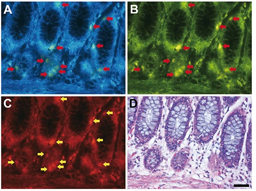
Figure 1.

Fluorescence imaging of formalin-fixed paraffin-embedded human colon tissue sections. A-C) Arrows indicate fluorescence signals; scale bar: 50 µm. The same section was subjected to fluorescence imaging through UV filter (A), blue filter (B) and green filter (C) after deparaffinization, and then, the section was stained with hematoxylin and eosin (D).
