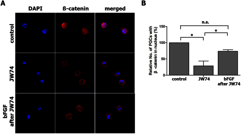
Figure 4. Activation and stabilization of β-catenin by bFGF treatment of chicken PGCs (SNUhp26).
(A) Immunostaining for β-catenin showed that the JW74 treatment caused the degradation of β-catenin in both the nucleus and the cytoplasm in cultured chicken PGCs. When bFGF was added followed by JW74 treatment, β-catenin translocated into the nucleus from the membrane. (B) The relative number of PGCs with β-catenin located in the nucleus was counted after each treatment. *P < 0.05. n.s.; not significant.

