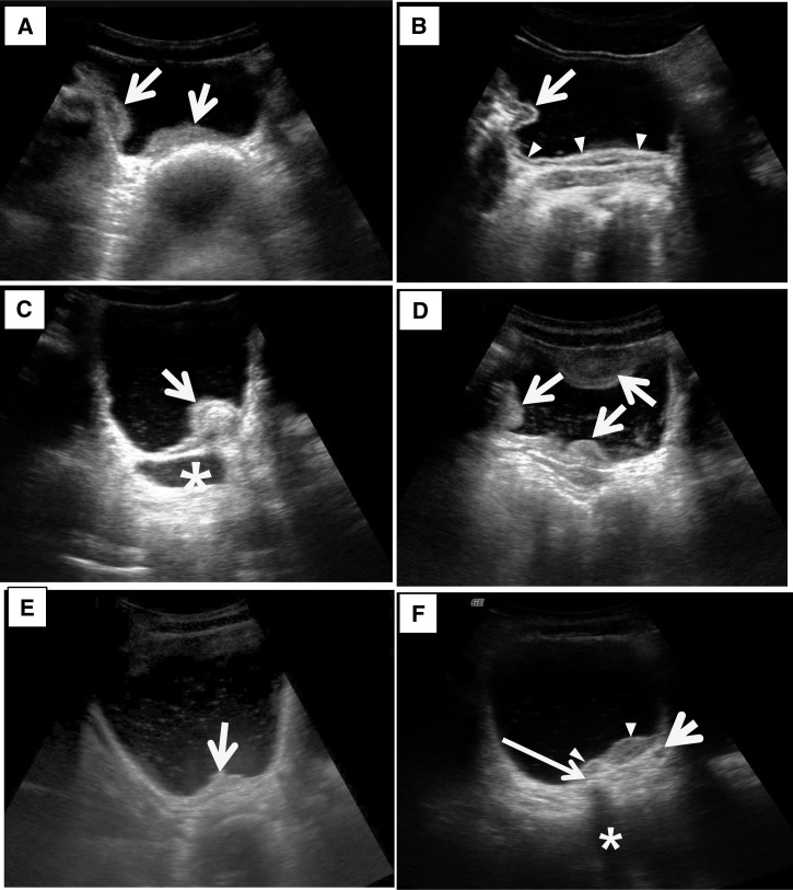
Figure 2.
Ultrasonography images of the urinary bladder with urogenital schistosomiasis. (A) Thickening of bladder wall. US image of the bladder in transverse plane shows marked thickening of the right lateral and posterior wall of the bladder (arrows). (B) Thickening of bladder wall. US image of the bladder in transverse plane shows diffuse thickening of the bladder wall (arrowheads) and a mass-like lesion (arrow) in the right lateral wall. Note that echogenic line of bladder mucosa is well preserved. (C) Bladder mass. US image of the bladder in oblique longitudinal plane shows a mass (arrow) in the posterior wall of the bladder. Note that echogenic line of the bladder mucosa is preserved indicating that the mass is in submucosal location. Also note that distal ureter is markedly dilated (asterisk) and there are fine scattered echoes (echogenic snow) in the dependent portion of the bladder. (D) Multifocal thickening of the bladder wall and echogenic snow in the lumen. US image of the bladder in transverse plane shows multifocal thickening of the bladder wall (arrows) and scattered echoes in the bladder lumen. (E) Focal thickening of the bladder wall and echogenic snow in the lumen. US image of the bladder in transverse plane shows focal thickening of the posterior wall of the bladder (arrow) and scattered echoes in the bladder lumen. (F) Focal thickening and probable calcification of the bladder wall and dilated ureter. US image of the bladder in transverse plane shows focal thickening of the posterior wall of the bladder (arrowheads). Note focal echogenic lesion (thin long arrow) with posterior sonic shadowing (asterisk) suggesting calcification of the bladder wall. Also note slightly dilated left distal ureter (short arrow).

