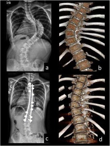
Fig. 2.

Preoperative (a, b) and postoperative (c, d: 1 week postoperatively) full-length standing posteroanterior radiographs and 3D reconstructed CT images in the main thoracic curve of an 11-year-old female patient (Patient No. 6 in Table 1). The 3D-CT images demonstrated that the opening of disc spaces in the apical region was observed at the concave side
