Figure 2.
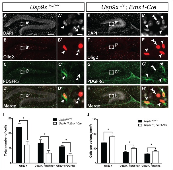
Increased density of oligodendrocytes in the dentate gyrus of Usp9x−/Y; Emx1-Cre mice. Co-immunofluorescence labeling and confocal microscopy was performed on hippocampal sections of Usp9xloxP/Y (A–D) and Usp9x−/Y; Emx1-Cre (E–H) at P14. Cell nuclei were labeled with DAPI (A, E). Oligodendrocyte precursors were defined as cells expressing both Olig2 (red in (B)and F) and PDGFRα (green in C and G). Mature oligodendrocytes were defined as cells that only expressed Olig2. The merged panels are shown in (D, H). The insets reveal a higher magnification view of the boxed region showing oligodendrocyte precursor cells (arrowheads) and mature oligodendrocytes (double arrowheads). Quantification of labeled cells was performed within the hilar region of the dentate gyrus. Total numbers of Olig2+ oligodendrocytes, including Olig2+/PDGRFα+ precursors and Olig2+/PDGRFα− mature oligodendrocytes in the mutant mice were significantly reduced compared to controls (I). Normalized cell counts relative to the volume of the hilar region revealed a significant increase in oligodendrocytic cells per mm3 within the mutant compared to controls, including elevated numbers of oligodendrocyte precursors and mature oligodendrocytes (J). *p < 0.05, t-test. Scale bar in (A): (A-H) – 150 µm; (A’): (A’-H’) −10 µm.

