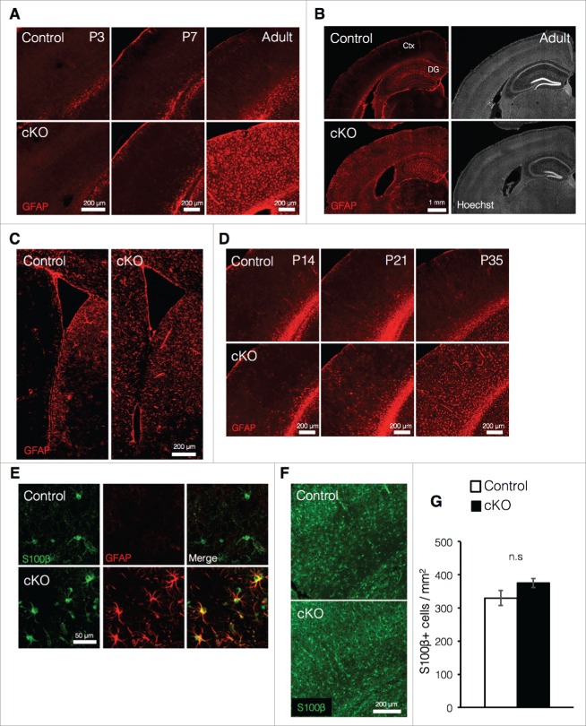
Figure 2.
Prenatal deletion of Dnmt1 increases expression of GFAP in astrocytes (A) Representative immunofluorescence images of GFAP (red) in the cortex of coronal brain sections from control and cKO mice at various time points. (B) GFAP immunostaining images (red, left panels) of representative cortical neurons in coronal adult brain sections from control and cKO mice. The nucleus was stained with Hoechst (gray, right panels). Ctx: cortex. (C) GFAP immunostaining images (red) of representative regions including the SVZ in coronal adult brain sections from control and cKO mice. (D) Representative GFAP immunofluorescence images (red) of cortical neurons in coronal brain sections from control and cKO mice at various time points. (E) GFAP (red) and S100β (green) immunostaining images of representative cortical astrocytes in coronal brain sections from adult control and cKO mice. (F) Representative image of staining for S100β (green) in the cortex of coronal brain sections from adult control and cKO mice. (G) Quantification of S100β+ cells in the cortex in (F). (Control = 3, cKO = 3). Scale bars are indicated in each figure. Values represent mean ± SEM; n.s > 0.05. Student's t-test. GFAP: glial fibrillary acidic protein.

