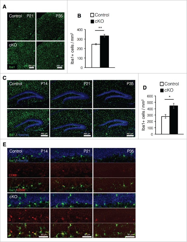
Figure 3.

Prenatal deletion of Dnmt1 increases microglia in the adult brain. (A) Representative Iba1 immunofluorescence images (green) in the cortex of coronal brain sections from control and cKO mice at various time points. (B) Quantification of IbaI+ cells in the cortex at P35. (Control = 3, cKO = 3). (C) Iba1 immunostaining image (green) of representative DG neurons in coronal brain sections from control and cKO mice at various time points. The nucleus was stained with Hoechst (blue). (D) Quantification of IbaI+ cells in the DG at P35. (Control = 3, cKO = 3) (E) Representative immunofluorescence images of Iba1 (green) and CD68 (red) in the DG of coronal adult brain sections from control and cKO mice at indicated time point. The nucleus was stained with Hoechst (blue). Scale bars are indicated in each figure. Values represent mean ± SEM; *P < 0.05, **P < 0.01. Student's t-test.
