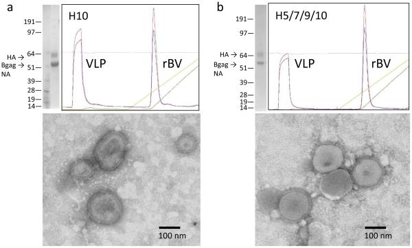
Fig. 1.
Preparation and characterization of mono-subtype H10 VLP (a) and quadri-subtype H5/H7/H9/H10 VLPs (b). Coomassie-stained is shown on the left upper panels, with molecular weight and locations of the HA, NA and Bgag are indicated. Anion exchange chromatogram is also shown in the upper panels. Bottom panels indicate the result of transmission electron microscopy of VLPs. Bar, 100nm.

