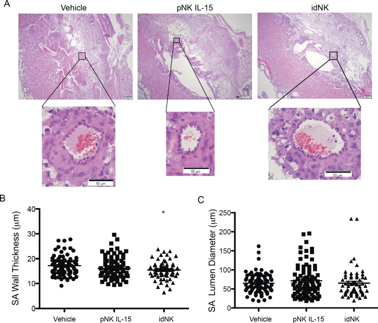
Fig 8. Effect of idNK injection on Rag2-/- γc-/- mice uterine spiral arteries.
(A) Photomicrograph of representative implantation sites at gd17 of mice injected on gd9 and gd12 with vehicle (PBS), pNK control cells cultured in IL-15, or idNK cells (upper images, scale bar 200μm). Illustrative decidual spiral arteries transversal sections are shown in higher magnification (lower images, scale bar 50μm). Spiral arteries muscular wall thickness (B) and lumen diameter (C) from implantation sites of mice receiving the indicated treatments. Each circle, square or triangle corresponds to an individual spiral artery transversal section. Mean is indicated with a line. Measurements were obtained from one or two Hematoxylin Eosin stained implantation site sections separated 42 microns apart, from each of 2 implantation sites per pregnant female. idNK and pNK IL-15 treated groups consisted each of 6 females, vehicle control group consisted of 7 females. The number of spiral artery transversal sections evaluated for each group were: Vehicle, 90; pNK IL-15, 85, idNK, 60. * p<0.05 between idNK and Vehicle by one way ANOVA with Bonferroni's Multiple Comparison Test.

