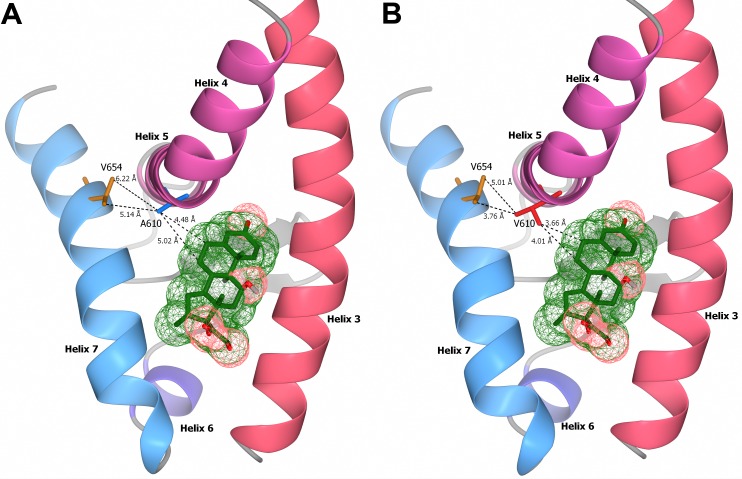
Fig 3. Stereogram of the interaction between glucocorticoid receptor ligand binding domain and dexamethasone.
Structural models of helix 3 to helix 7 of the ligand binding domain of the wild-type Ala610 (A) and the mutated Val610 (B) receptor variants are shown. The distances of the alternative alanine (blue) and valine (red) residue at amino acid position 610 with the two methyl carbon atoms of Val654, and the carbon C-6 and C-7 positions of the ligand dexamethasone (green), are indicated in angstroms (Å).

