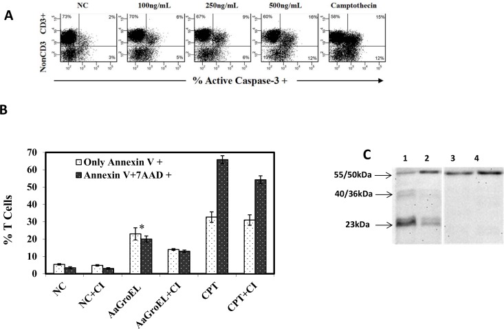
Fig 3. GroEL induces T cell apoptosis by activating caspases-3 and -8.
(A) Caspase-3 activation in T cells. PBMCs were cultured for 72 h with various AaGroEL doses. RPMI alone (NC) and camptothecin (CPT, 4 μM) were used as negative and positive controls, respectively. Cells were first labeled with anti-CD3 antibody. The cells were then fixed and permeabilized, and anti-active caspase-3 antibody was added, followed by analysis using flow cytometry. The representative flow data indicate the active-caspase-3 level (%) of T cells (CD3+) and non-CD3+ cells. (B) Caspase inhibition assay. PBMCs were incubated with a general caspase inhibitor (CI; Z-VAD-FMK) for 1 h at 37°C before antigenic stimulation. After this incubation, the PBMCs were cultured with AaGroEL (250 ng/mL) and camptothecin (CPT, 4 μM) for 72 h. At the end of the culture, the cells were labeled with anti-CD3 antibody, Annexin V and 7AAD and analyzed by flow cytometry. Error bars represent the standard deviation, and * indicates p<0.05. The data are representative of three experiments with different donors. (C) Caspase-8 activation. PBMCs were cultured with AaGroEL (250 ng/ml) protein for 72 h and 48 h (lanes 1 and 2, respectively). RPMI was used as a negative control (lanes 3 and 4). The cells were probed with anti-human caspase-8 antibody and analyzed by western blotting. The 55/50 kDa procaspase-8 was detected in all the samples (lanes 1 to 4). The cleaved 40/36-kDa (doublet) and 23-kDa caspase-8 bands were seen only in AaGroEL-stimulated cells at 72 h and 48 h, respectively.

