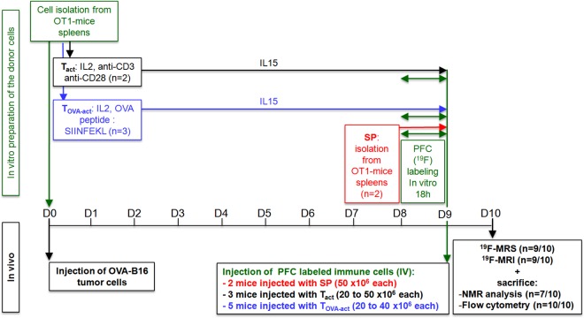
Fig 1. In-vivo protocol description.
Overview of the time scale of the different experimental procedures. At day 0 (D0) eight CD45.1 C57BL/6 mice received 106 B16-F10 melanoma cells by subcutaneous injection in order to induce a malignant melanoma. On the same day SP were prepared from OT-1 mice and two different protocols were applied to generate Tact or TOVA-act (as described in Materials and Methods, T cells isolation and activation section). At day 8 (D8), PFC was added in the cell culture medium for 18h in order to label SP, Tact and TOVA-act with 19F. Then, at day 9 (D9) the 19F-labeled cells were injected IV: 2 control mice (with no tumors) received 50 x 106 SP, 3 mice received 20 to 50 x 106 Tact and 5 mice received 20 to 40 x 106 TOVA-act. Finally, 9 mice were imaged at day 10 (D10; 1 Tact injected mouse was not imaged) and all mice were immediately sacrificed for subsequent analysis of the organs (liver, lungs, spleen and tumor) by flow cytometry (all mice) and high resolution in-vitro NMR spectroscopy (2 SP injected mice, 3 TOVA-act injected mice and 1 Tact injected mouse). The study protocol was performed in a total of 10 animals. In black: in-vivo part; in blue: cell preparation.

