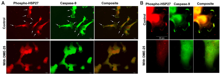
Figure 3.
(A) Co-staining phospho-HSP27 and caspase-9 in human lung cancer SKMES-1 cells. Control cells displayed high levels of staining of both phospho-HSP27 and caspase-9, both also showing a high degree of co-localisation (white arrows). However, the co-localisation was eliminated by DME25 (bottom images). (B) Images under a higher power showing focal adhesion (FAC) and pseudopodia within the SKMES-1 cells.

