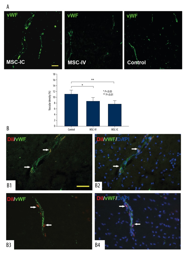
Figure 5.
Pro-angiogenic effect of UC-MSCs. (A) Blood vessels are recognized by vWF immunostaining (green); representative images show that the vascular density in the MSC-IC group is significantly higher than that in the MSC-IV and control groups. The vascular density is not different between the MSC-IV group and the control group. n=3 per group. (B) At 14 days after treatment, in the MSC-IV group, a population of CM-DiI-labeled UC-MSC cells (red) either located in the endothelium and expressed endothelial-specific marker vWF (B1, B2) or located in blood vessel walls (B3, B4). DAPI (blue) was used to identify the nucleus. Scale bars=50 μm.

