Figure 1.
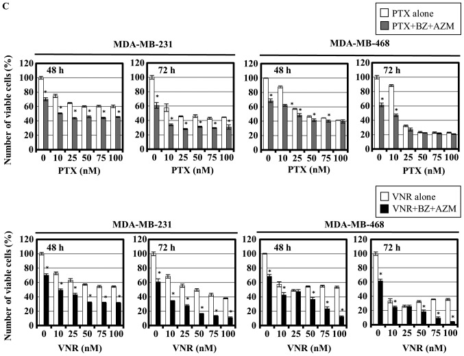
Cell growth inhibition and apoptosis induction of MDA-MB-231 and MDA-MB-468 cells after combined treatment using PTX or VNR and BZ in the presence or absence of AZM. (A) MDA-MB-231 and MDA-MB-468 cells were treated with BZ at various concentrations for 24, 48 and 72 h. The viable cell number was assessed using CellTiter Blue assay as described in Materials and methods. (B) MDA-MB-231 and MDA-MB-468 cells were treated with PTX or VNR at various concentrations with/without BZ (25 nM for MDA-MB-231 cells and 7.5 nM for MDA-MB-468 cells) for 48 and 72 h. *P<0.05; PTX vs. PTX+BZ, VNR vs. VNR+BZ. (C) MDA-MB-231 and MDA-MB-468 cells were treated with PTX or VNR in the presence of BZ (25 and 5 nM) and AZM (50 μM) for 48 and 72 h. *P<0.05; PTX vs. PTX+BZ+AZM, VNR vs. VNR+BZ+AZM. (D) MDA-MB-231 cells were treated with BZ (25 nM), PTX (50 nM), or VNR (50 nM) alone or a combination of BZ and PTX, or BZ and VNR for 24 h. Flow cytometric analysis with Annexin V/PI double staining was performed. Numbers indicate the percentage of the cells in each area to the cells in whole gating.


