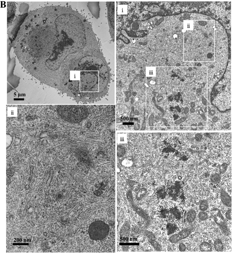
Figure 2.
Aggresome formation after treatment using BZ in MDA-MB-231 cells. (A) After 24-h treatment using BZ at 25 nM, immunocytochemistry was performed using anti-vimentin mAb, anti-ubiquitin mAb, and anti-p62 mAb. DAPI staining indicates the position of the nucleus (blue). Dotted white line represents cell outline. Scale bar 10 μm. (B) Electron microscopy: MDA-MB-231 cells were treated with BZ (25 nM) for 24 h.


