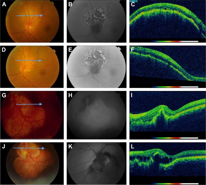
Figure 3.
Color fundus photographs, FAF images, and OCT scans of patients with breast cancer metastasis and choroidal osteoma.
Notes: Images from a female aged 72 with breast cancer metastasis in her left eye (A–F): (A and D) Color fundus photos show nonpigmented tumor (A) and 3 months after cytostatic agents choroidal mass reduction with partial pigmentation (D). (B and E) FAF images show a patchy pattern characterized by distinct areas of increased autofluorescence between areas of normal autofluorescence (B), and 3 months after treatment the FAF is unchanged (E). (C and F) OCT scans before (C) and after treatment (F) show minimal intra- and subretinal fluid. Images from a female aged 30 with choroidal osteoma in her left eye (G–L). (G and J) Color fundus photos show orange-yellow inferotemporal tumor in contact with the optic nerve (G) and minimal progress of the tumor 10 years later (J). (H) Indocyanine green angiography, late phase shows diffuse hyperfluorescence due to staining. (K) FAF image shows mostly hypoautofluorescence due to RPE atrophy. (I and L) OCT scans show RPE detachment (I) and increased subretinal fluid 10 years later (L). The arrows at the color fundus photographs (A, D, G, J) correspond to the B-scan lines of the OCT images.
Abbreviations: FAF, fundus autofluorescence; OCT, optical coherence tomography; RPE, retinal pigment epithelium.

