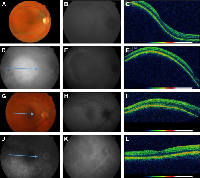
Figure 4.
Color fundus photographs, FAF images, and OCT scans of patients with choroidal hemangioma and choroidal lymphoma metastasis.
Notes: Images from a female aged 42 with a choroidal hemangioma in her right eye (A–F). (A) Color fundus photo shows an orange-pink circumscribed tumor. (B) FAF image shows isoautofluorescence over the tumor. (C and F) OCT scans of the tumor reveal the abruptly elevated choroidal mass without intra- or subretinal fluid. (D and E) ICG in the midphase shows homogeneous hyperfluorescence of the tumor (D) and in the late phase dye washout with peripheral leakage (E). Images from a female aged 34 with a choroidal metastasis of systemic lymphoma in her right eye (G–L). (G) Color fundus photo shows a nonpigmented choroidal mass. (H) FAF image shows isoautofluorescence of the tumor. (I) OCT scan reveals the abruptly elevated choroidal mass. (J) FA, late phase shows minimal hyperfluorescence. (K) ICG angiography, late phase shows hypoautofluorescence of the tumor. (L) Normalized OCT scan 1 month after treatment with cytostatic agents. The arrows at ICG image (D), the color fundus photograph (G), and FA image (J) correspond to the B-scan lines of the OCT images.
Abbreviations: FA, fluorescein angiography; FAF, fundus autofluorescence; ICG, indocyanine green angiography; OCT, optical coherence tomography.

