FIGURE 1:
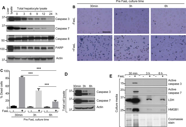
Caspase 3 and 7 levels decrease rapidly with parallel relative resistance to apoptosis during short-term mouse hepatocyte culture. (A) Hepatocytes were isolated from FVB/N mice and cultured for the indicated times after plating onto collagen-coated plates. Total cell lysates were analyzed by blotting, using antibodies to the indicated proteins. A total liver lysate (lane 1) is included to show in situ whole-liver caspase levels, and an actin blot is included as a loading control. (B) Hepatocytes were isolated from FVB/N mice, followed by treatment with FasL (0.5 μg/ml, 6 h) in the presence of trypan blue (0.04%) after 30 min, 3 h, or 6 h of cell culture (see Supplemental Figure S1 for culture conditions). Scale bar, 200 μm. (C) Quantification of percentage of dead cells from the data in B, based on trypan blue uptake. A repeat experiment showed essentially identical findings. ***p < 0.001. (D) Total cell lysates were prepared from the hepatocytes used in B (just before the addition of FasL), followed by blotting for intact caspases 3 and 7. (E) Culture medium from a parallel experiment to that shown in B was concentrated and then analyzed by blotting to assess the release of the indicated proteins. A Coomassie stain of the concentrated culture medium is included and shows the limited amount of protein released when cells are cultured for 3 and 6 h before the addition of FasL (lanes 3 and 5).

