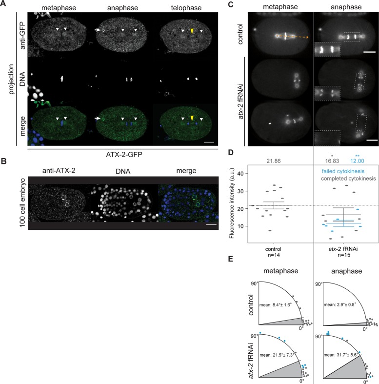
FIGURE 2:
ATX-2 localizes to mitotic structures and is required for spindle orientation and midzone dynamics. (A) Fixed ATX-2–GFP embryos in metaphase, anaphase, and telophase and stained with anti-GFP antibody (green) and DAPI (blue). ATX-2–GFP localized to cytoplasmic puncta, 2-μm spherical aggregates (white arrows), centrosomes (white arrowheads), and the spindle midzone (yellow arrowheads). Images are average projections of 0.1-μm Z-stacks spanning 1.5 μm. Scale bar, 10 μm. (B) Fixed 100-cell stage N2 embryo stained with anti–ATX-2 antibody (green) and DAPI (blue). ATX-2 localized to cytoplasmic puncta and Z2 and Z3 cells. Image is a single focal plane. Scale bar, 10 μm. (C) Microtubule dynamics in control and atx-2 fRNAi–treated embryos coexpressing TBB-2–GFP and GFP–HIS-11 in metaphase and anaphase. Insets, 1.5×-magnified view of spindle midzone microtubules in dashed boxes. Control embryos contain discrete midzone microtubules, whereas atx-2 fRNAi–treated embryos show faint or no visible midzone microtubules. Orange arrow shows angle measurement relative to horizontal (0º). (D) Quantification of TBB-2–GFP fluorescence intensity in control and atx-2 fRNAi–treated embryos. Each point represents a fluorescence intensity measurement from a single embryo. Gray points represent embryos in which cytokinesis successfully completed; blue points represent embryos in which cytokinesis failed to complete. Reduced TBB-2–GFP fluorescence intensity at the spindle midzone was observed in ATX-2–depleted embryos. Solid lines denote the mean. Results are mean ± SEM. Asterisks represent level of significance for compared data. *p < 0.05, **p < 0.01 (Welch’s t test). (E) Quantification of spindle displacement from 0º measured at metaphase and anaphase in control (n = 14) and atx-2 fRNAi–treated (n = 15) embryos. Gray points represent embryos in which cytokinesis successfully completed; blue dots represent embryos in which cytokinesis failed to complete. In control embryos, the spindle is oriented along the anterior–posterior axis, with average displacement varying between 3 and 8º. In atx-2 fRNAi–treated embryos, the average displacement increases to between 21 and 32º. Extreme spindle orientation defects correlate with cytokinesis defects in atx-2 fRNAi–treated embryos.

