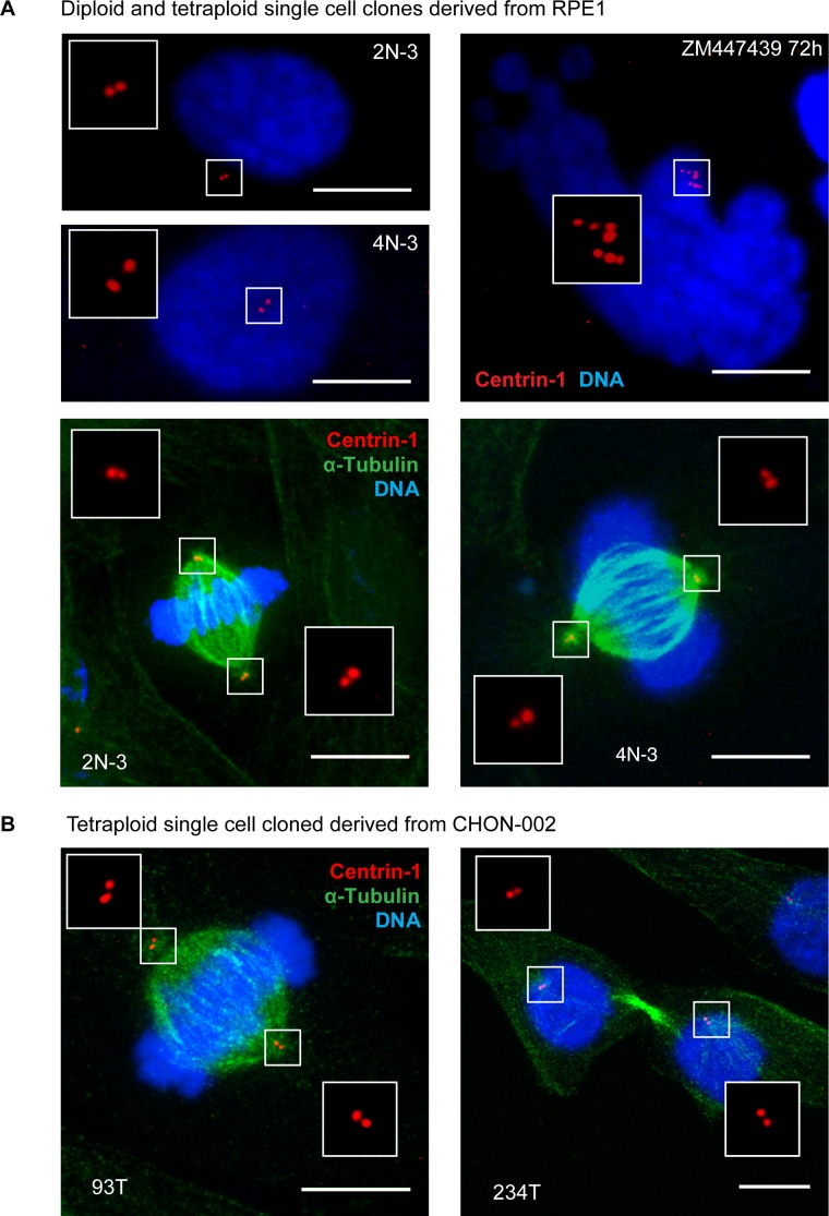
FIGURE 6:
Established proliferating tetraploid cells have normal number of centrioles. (A) Immunofluorescence of a centriole component centrin-1 (red). Top left, diploid and established tetraploid cells in interphase. Note two centrin-1 dots in both cells. Top right, acute polyploid cell in interphase. This cell was treated with Aurora kinase inhibitor ZM447439. It displays characteristic nuclear morphology and has multiple centrioles. Bottom, diploid and proliferating tetraploid cells have equal centrosome number and form bipolar mitotic spindles in metaphase. DNA was counterstained with DAPI (blue); α-tubulin was labeled by immunostaining (green). At least 30 cells were examined for every image. Maximum projections of representative confocal stacks. (B) Centrin-1 labeling of proliferating tetraploid CHON-002 single-cell clones. Immunofluorescence labeling of centrin-1 (red) and α-tubulin (green) of proliferating tetraploid single-cell clones designated 93T (metaphase cell) and 234T (telophase cell). Nuclei were counterstained with DAPI (blue). Maximum projections of confocal stacks. Like proliferating tetraploid RPE1, established tetraploid cells derived from CHON-002 have centrosome number equal to diploid and form bipolar mitotic spindles. Bar, 10 μm.

