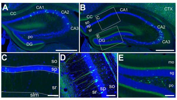
Figure 3. EGFP-immunoreactive cells in the hippocampus.
A) Low-resolution immunofluorescence overview of EGFP-immunoreactivity in a relatively anterior section of the hippocampus. EGFP-labeled cell bodies (green) are found primarily in the pyramidal layer of CA1 as well as in the polymorph layer (po) of the dentate gyrus. Some cell bodies are intensely labeled in CA2 and CA3, as well, though in smaller proportion to CA1 Diffuse and fibrous staining is found in the CC, stratum radiatum (sr), particularly in the ventral portion that abuts the dentate gyrus, as well as a tight band in the molecular layer of the dentate gyrus (DG). Nissl staining (blue) provides the anatomical structure. Scale bar = 500μm. B) Another cross section of the hippocampus, representing a more central portion than in (A). Here, there is denser labeling of cells in the CA2 and CA3 region. A similar pattern of staining to (A) is found in other subregions. C) High-resolution inset of the CA1 region defined in (B). Some cell bodies and processes with neural morphology are stained in this region, but sparsely at this position along the rostro-caudal axis. Scale bar = 100μm. D) Confocal image of the CA3 region of the hippocampus. Cells with a pyramidal morphology are labeled in the pyramidal layer (sp), with processes extending out to the stratum oriens (so) and sr. E) High-resolution inset of the DG defined in (B). Very dense fibrous staining is observed in the medial perforant pathway in the molecular layer (mo) abutting the granule layer (sg). Additionally, large cells within the polymorph layer are intensely stained for EGFP. Scale bar = 200μm.

