
Figure 8. EGFP immunoreactivity in the cervical and thoracic spinal cord.
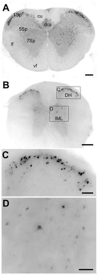
A) Low resolution image of a cross-section of the cervical spinal cord. The gray matter of the spinal cord is diffusely labeled for EGFP, as is the dorsal corticospinal tract (dcs). Intense labeling of cell bodies is observed in lamina 1, with more scattered cell labeling in laminae 5 and 7. Scale bar = 200μm. B) Low-resolution image of a cross-section of thoracic spinal cord. There is a similar pattern of EGFP immunoreactivity to that in (A). Scale bar = 200μm. C) High-resolution inset of the dorsal horn area defined in (B). Many cells in this area are intensely stained. Scale bar = 50μm. D) High-resolution inset of the intermediolateral column (IML) showing dispersed neurons and processes stained for GFP. Scale bar = 50μm.