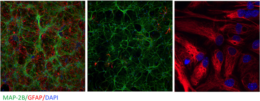
Figure 3.
Representative photomicrographs of cultures used for in vitro studies. Enriched neurons (left panel), enriched astrocytes (middle panel), and neuron–astrocyte co-cultures (right panel) immunostained for the dendritic biomarker MAP-2b (green), the astrocyte biomarker GFAP (right), and the nuclear stain DAPI (blue). Magnification differs between images.

