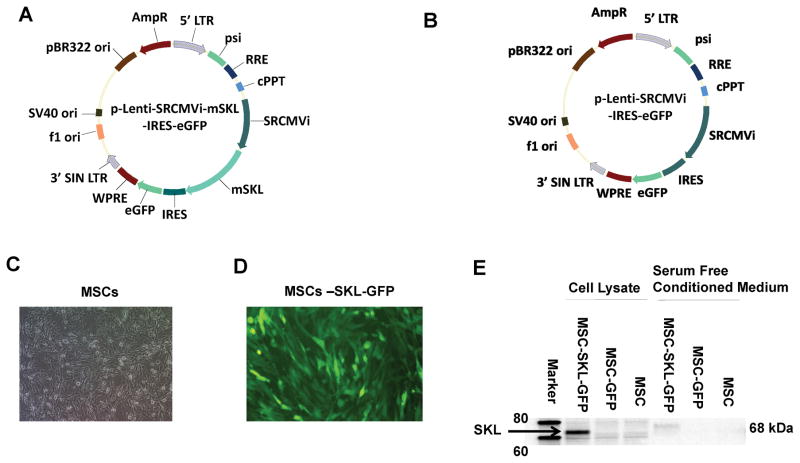
Figure 1. Transfection of mesenchymal stem cells (MSCs) with the mouse secreted Klotho (mSKL) gene.
A) Map of lentiviral vector expressing the mSKL and eGFP genes. LTR, long terminal repeat; psi, ψ domain; RRE, Rev responsive element; cPPT, central polypurine tract; SRCMVi, a CMV promoter plus enhancers from RSV and SV40 promoters and a human β-globin intron; mSKL, mouse secreted Klotho gene; IRES, internal ribosome entry site; eGFP, enhanced green fluorescent protein gene; WPRE, woodchuck hepatitis virus post-transcriptional regulatory element; SIN, self-inactivating LTR as a result of the deletion of the U3 promoter. B) Map of the control lentiviral vector expressing eGFP only. C) Phase-contrast image of MSCs in culture. D) eGFP expression in Lenti-SKL-GFP-transduced MSCs. E) Western blot analysis of SKL protein expression in cell lysates and conditioned medium from transfected and non-transfected MSCs.

