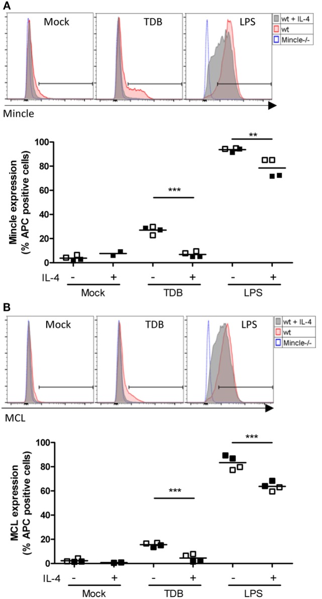
Figure 4.

Regulation of cell surface levels of Mincle and Mcl in murine macrophages by IL-4. Bone marrow-derived macrophages were analyzed for Mincle (A) and Mcl (B) surface expression 16 h after stimulation as indicated by using anti-Mincle (4A9) or anti-Mcl (3A4) primary antibodies followed by staining with APC-conjugated secondary antibody. (A) Histograms show Mincle surface expression in C57BL/6 BMMs (wt) stimulated with TDB or LPS or left untreated (Mock) in presence (gray filled) or absence (red filled) of IL-4 as indicated. Mincle−/− BMMs (blue dotted) were stimulated with TDB or LPS or left untreated and used as negative control. Histograms are from one representative experiment. Scatter plot shows Mincle expression depicted as percentage of APC-positive cells from C57BL/6 BMMs after stimulation with TDB or LPS in presence or absence of IL-4 or left untreated (Mock). Data are depicted from two independent experiments performed in duplicates (filled symbols: exp. 1, open symbols: exp. 2). (B) Histograms and scatter plot show Mcl surface expression, respectively. Cells were treated as described in (A). Statistical significance was tested using an unpaired t-test. **p < 0.01, ***p < 0.001.
