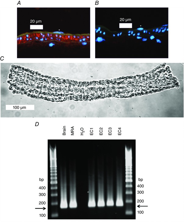
Figure 7. Expression of CaV3.2 at the mRNA and protein level in young mouse SMAs .

A, mouse SMA (young) stained with primary antibody against CaV3.2 (red colour). The internal and external elastic laminas are visible as faint green bands using the inherent autofluorescence of the tissue. Nuclei are stained blue with 4′,6‐diamidino‐2‐phenylindole. An endothelial cell nucleus is marked with an asterisk in the vessel lumen. Clear CaV3.2‐specific red staining is present in the media layer and faint red staining is visible in the intima layer on the luminal side of the green internal elastic lamina. B, negative peptide pre‐absorption control staining for CaV3.2 primary antibody of mouse mesenteric artery (young). Note the absence of a positive red signal. An endothelial cell nucleus is marked with an asterisk in the vessel lumen. C, image of isolated endothelial tube from a young mouse SMA. The image was acquired with a Axiovert S‐100 microscope (Carl Zeiss, Oberkochen, Germany) through a Neofluar 20× objective using a USB camera and Myoview II software. D, agarose gel showing electrophoresis of Cacna1H qPCR products from mouse brain (n = 1), mouse whole SMA (n = 1) and SMA endothelial tubes isolated from four young WT mice (EC1–EC4). Horizontal arrows depict the product size corresponding to Cacna1H (169 bp). Note the similar product size in brain, whole SMA and EC tubes.
