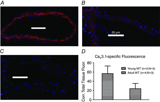
Figure 8. Age‐dependent intensity of CaV3.1‐specific staining in mouse SMAs .

Immunostaining of the CaV3.1 T‐type isoform (red colour) in young (A) vs. mature adult (B) WT mouse mesenteric arteries, and in staining without primary antibody (C). Note the lower fluorescence intensity visible in the mature adult artery compared to the young artery. Images were acquired using same settings of microscope, camera and software. Quantitation of background‐corrected total tissue fluorescence in young vs. mature adult arteries using ImageJ (D).
