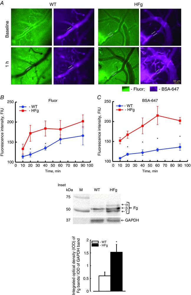
Figure 1. Hyperfibrinogenaemia‐induced leakage of mouse pial venules .

A dual‐tracer probing method was used to define the prevailing role of transcellular vs. paracellular transport in mouse brain pial venules. A, examples of images of cerebral vessels recorded immediately (baseline, upper row) and 1 h after (lower row) infusion of Fluor (green) and BSA‐647 (violet) dyes in wild‐type (WT, left) and hyperfibrinogenic (HFg, right) mice. Cerebrovascular permeability was assessed by comparison of ratios of fluorescence intensities of dyes measured along the line profile probe (shown as a yellow line segment) outside to that inside of the vessel. Summaries of changes in fluorescence intensity ratios of Fluor (B) and BSA‐647 tracers (C). * P < 0.05 vs. WT; n = 8 for all groups. Inset: top, Western blot analysis of plasma samples from WT and HFg mice shows content of Fg (α, β and γ chains); bottom, summary of ratios of integrated optical density (IOD) of Fg bands to IOD of respective GAPDH (used as a loading control) bands. * P < 0.05 vs. WT; n = 6 for all groups. [Colour figure can be viewed at wileyonlinelibrary.com]
