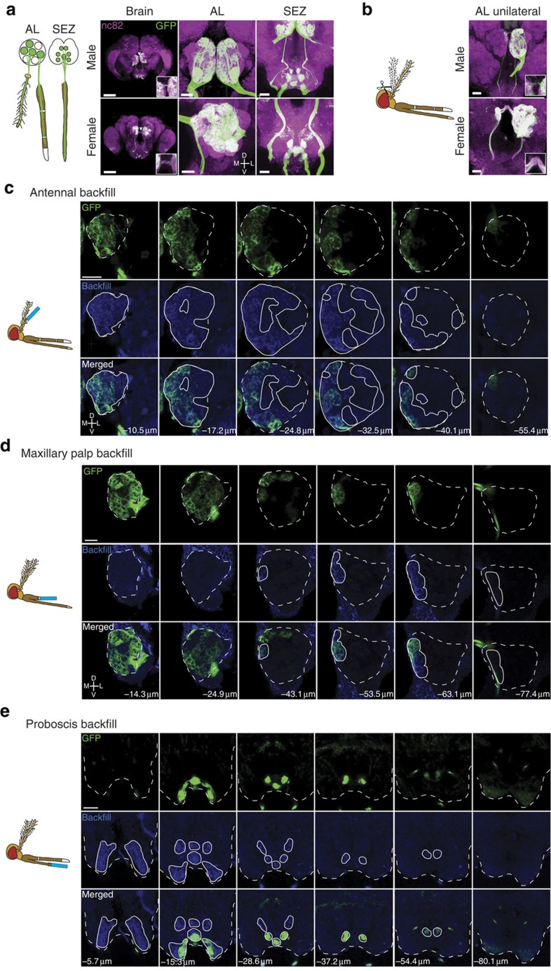
Figure 3. Orco+ olfactory neurons target two sensory brain regions.
(a) Orco+ receptor neurons send projections to the AL and SEZ of the brain, as detected in Orco-QF2, QUAS-mCD8:GFP animals by anti-GFP antibody labelling. Left column shows an overall image of the brain (scale bar, 100 μm). Middle and right columns show the AL and SEZ at higher magnifications (scale bar, 20 μm). Confocal sections are maximum z-projections. Arrowhead points to commissure. Inset shows commissure at higher magnification. (b) Shown are brains of male and female Orco-QF2, QUAS-mCD8:GFP mosquitoes in which the right antennae had been ablated (as schematized in the cartoon) 5 days before brain dissections. The ALs retain ipsilateral Orco+ innervation from the unablated left antennae and bilateral innervation from the intact maxillary palps. Inset shows commissure at higher magnification. Scale bars, 20 μm (c–e). Antennae (c), maxillary palps (d) or proboscis (e) of Orco-QF2, QUAS-mCD8:GFP mosquitoes were cut to ∼1/3 of their length (as depicted in the cartoons) and backfilled by neurobiotin that is incorporated into the membranes of severed neurons. The neurobiotin labelling (blue) of the brains, together with the GFP labelling (green), establishes the origin of Orco+ receptor neurons (antennal, maxillary palp or proboscis) that innervate the AL and SEZ. Backfills originating from the antenna innervated only the ipsilateral AL. Backfills originating from the maxillary palps innerved both ipsilateral and contralateral ALs. Backfills originating from the proboscis innervated only the SEZ brain region. The numbers indicate the distance in micrometres from the most anterior confocal section. Dashed white lines outline the AL/SEZ. Solid white lines outline the area labelled by the neurobiotin backfill (blue signal). Scale bars, 20 μm.

