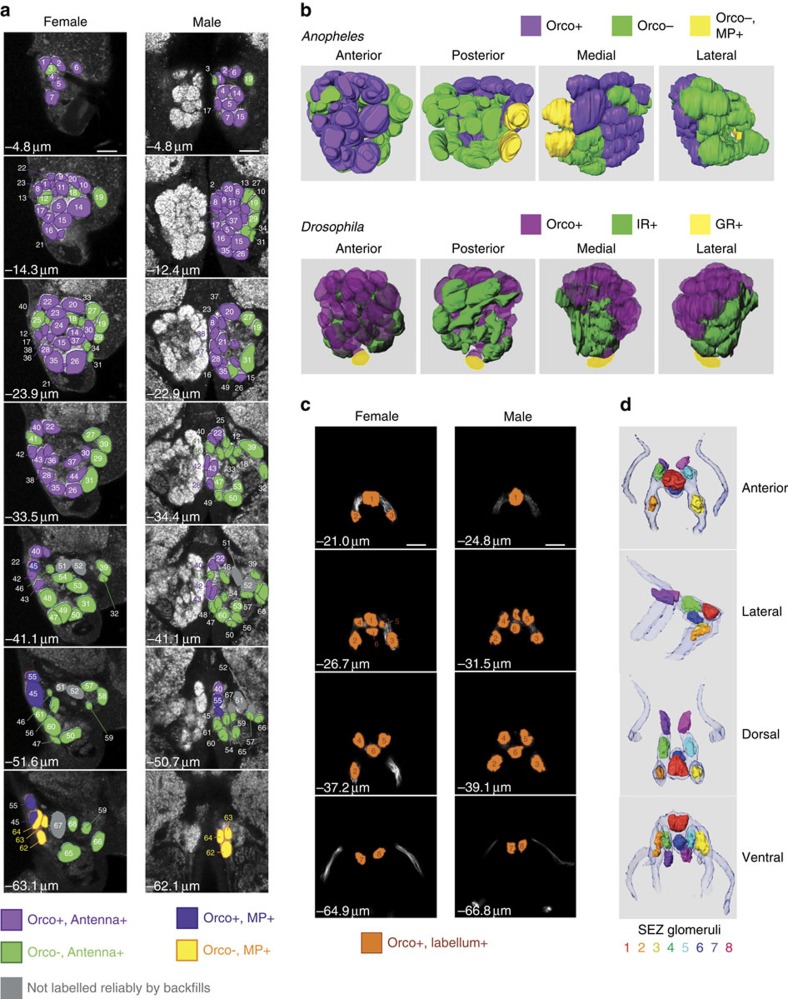
Figure 4. Reconstruction of the mosquito AL and SEZ based on Orco expression and olfactory tissue of origin.
(a) Confocal Z-stacks of female (left) and male (right) ALs, shown at the same magnification (most anterior—top row, most posterior—bottom row). Glomeruli were outlined manually, and GFP and neurobiotin backfill signals were used to assign each glomerulus to one of five groups: Orco+ antennal glomeruli (purple), Orco− antennal glomeruli (green), Orco+ maxillary palp glomeruli (blue), Orco− maxillary palp glomeruli (yellow) and glomeruli that were not labelled by backfills (grey). Glomeruli were numbered starting at the most anterior section. Scale bars, 20 μm. Also see Supplementary Table 5. (b) A. gambiae and D. melanogaster ALs show similarities in the arrangement of glomeruli targeted by Orco+ (purple) and Orco− neurons. Orco− neurons originating from the mosquito antennae are likely to be IR-expressing neurons (IR, green) and Orco− neurons from the maxillary palps are likely to be GR-expressing neurons (GR, yellow)22. The Drosophila AL is reprinted with permission from ref. 76 with minor modifications. (c) Confocal Z-stacks of female (left) and male (right) SEZ, shown at the same magnification (most anterior, top row; most posterior, bottom row). Glomerular-like structures were outlined manually using the GFP signal. Glomerular structures were numbered starting at the most anterior section. Scale bars, 20 μm. (d) Three-dimensional modelling of olfactory neuron targeting in the SEZ. The Orco-QF2-labelled nerve bundle from the labella is shown for reference.

