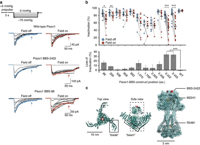
Figure 2. Effect of magnetic pulling force on Piezo1 inactivation.
(a) Representative current recordings of wild-type Piezo1, construct BBS-2422 and construct BBS-86 on pressure-clamp stimulation from 0 to −70 mm Hg (above) in the absence and presence of a magnetic field. Arrows denote inactivated current after 150 ms. Traces highlighted in bold represent −60 mm Hg step and corresponding current (blue, field off; red, field on). (b) Inactivation at −60 mm Hg (or −110 mm Hg for BBS-1758; top panel) for individual experiments and averages of constructs labelled with nanoparticles before (blue) and during (red) application of magnetic force (n=8–21 cells, 3–7 transfections; *P<0.01 **P<0.001, ***P<0.0001, paired t-test). Average loss of inactivation (bottom panel) for data above (***P<0.0001, one-way ANOVA and Tukey's multiple comparison and NP multiple comparison). Error bars are s.e.m. (c) Top and side views (left and middle panels) of MmPiezo1 cryo-EM study (PDB 3JAC) with approximate location of aa. position 2,422 highlighted in red. Previously named ‘blade' and ‘beam' are boxed. Predicted pore region (right) with overlaid electron density, highlighting 2,422 position in red and alignment of human Piezo1 inactivation point mutations in yellow (M2242 and R2481 in mouse).

