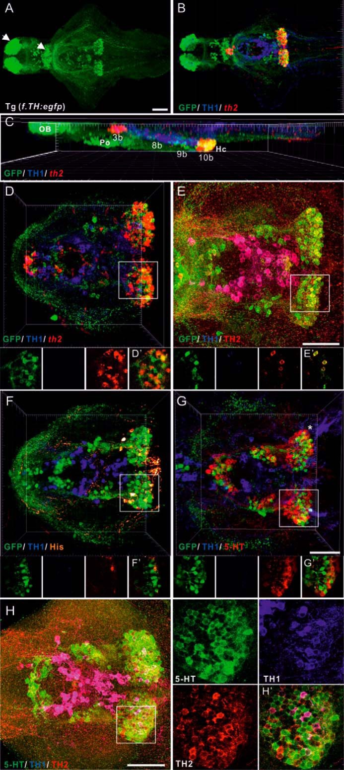
FIGURE 1.

Multiple labeling of catecholaminergic, histaminergic, and serotoninergic neurons and GFP distribution in 5-dpf brains of the Tg(f.TH:egfp) transgenic line and Turku WT. A, ventral view of GFP distribution in a 5-dpf brain. The specimens initially hybridized (ISH) with th2 antisense riboprobes were processed for double immunostaining with chicken GFP and mouse TH1 antibodies (shown in B–D). B, ventral view of triple staining showing GFP-ir in green, TH1-ir in blue, and the th2 mRNA expression pattern in red. C, lateral view of B. The distribution patterns in the diencephalic and hypothalamic regions of GFP and TH1 with TH2, histamine (His), or 5-HT are shown in E–G, respectively. A triple immunostaining image with anti-TH1, anti-TH2, and anti-5HT on a 5-dpf brain is shown in H and H'. Larger magnification and single section images of group 10b (white rectangle) in D–H are shown in D'–H', respectively. The GFP-ir signal is shown in green, TH1-ir in blue, and th2-ish, TH2-ir, His-ir, and 5-HT-ir in red in the transgenic line (D–G). 5-HT-ir is shown in green in H. White arrows indicate GFP-positive but th2/TH2-negative populations. Cells labeled with both TH1 and th2 are shown in magenta. Cells labeled with both GFP and TH2 are shown in yellow. OB, olfactory bulb. 3b, preoptic group (Po); 8b, paraventricular organ; 9b, nucleus of lateral recess; 10b, caudal hypothalamus (Hc). TH2 group numbers are based on Ref. 13. Scale bars = 50 μm.
