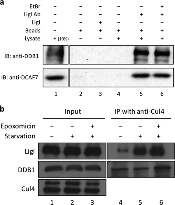
FIGURE 6.

Association of LigI with the Cul4-DDB1-DCAF7 complex. a, lane 1, 293T cell lysate, 5 μg (10% input). Lane 2, protein A/G beads only. Lane 3, protein A/G beads plus LigI. Lanes 4–6, 293T cell lysate incubated with: protein A/G beads (lane 4); LigI antibody (LigI Ab) followed by protein A/G beads (lane 5); LigI antibody plus 10 μg/ml ethidium bromide followed by protein A/G beads (lane 6). DDB1 and DCAF7 proteins were detected by immunoblotting (IB). b, input lysates (5 μg, 10% of input) and Cul4 immunoprecipitates (IP) from GM00847 cells that were: untreated (lanes 1 and 4); serum-starved for 8 h (lanes 2 and 5); serum-starved for 8 h in the presence of 0.2 μm epoxomicin for 8 h (lanes 3 and 6). Cul4, DDB1, and LigI proteins were detected by immunoblotting.
