FIGURE 3.
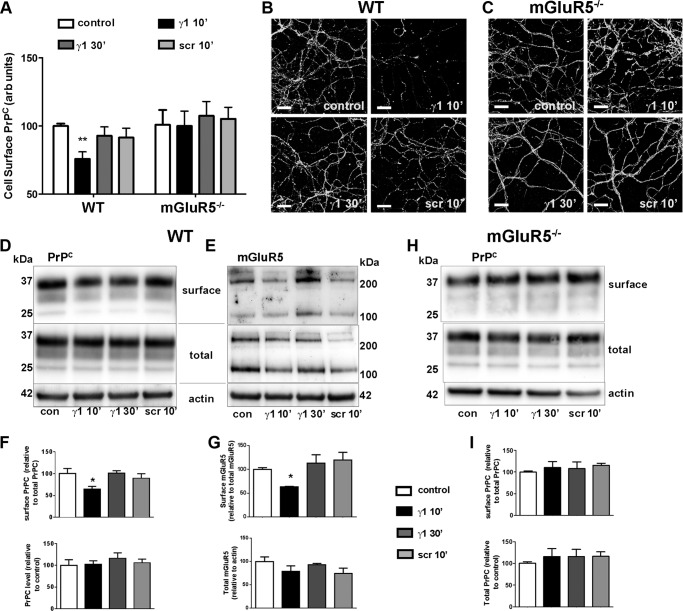
Ln-γ1-induced transient internalization of PrPC and mGluR5 in primary neurons. A–C, WT (A and B) and mGluR5−/− (A and C) hippocampal neuronal cultures were treated with Ln-γ1 for 10 or 30 min, or with Ln-SCR (Ln-γ1 scrambled peptide: IRADIEIKID), and the neuronal cell surface was immunostained with 8H4 antibody. Quantification of surface PrPC was done for eight random fields of view for each condition (A). Primary neuronal cultures were prepared from at least five embryos of each genotype. arb units, arbitrary units. B and C, representative images of WT (B) and mGluR5−/− (C) cultures, corresponding to each treatment. Scale bar = 20 μm. D–I, primary cortical neurons were treated with Ln-γ1 for 10 or 30 min, or with Ln-SCR, after which the surface proteins were biotinylated. Levels of surface PrPC (D and F) and mGluR5 (monomer + dimer, E and G) in WT cultures, and of surface PrPC in mGluR5−/− cultures (H and I), were quantified as the ratio of biotinylated protein to the total protein levels on Western blot (D, E, and H), and then compared using GraphPad 5.0 software (F, G, and I). con, control. Error bars indicate mean ± S.E. *, p < 0.05.

