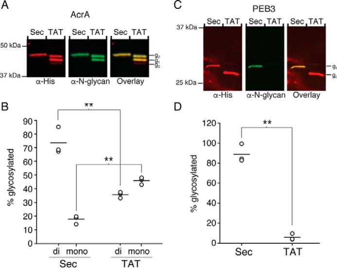
FIGURE 5.

Glycosylation of protein substrates in C. jejuni. Representative Western blot results of periplasmically-localized AcrA (A) and PEB3 (C) translocated via the Sec or TAT pathway in C. jejuni (n = 3) are shown. Total AcrA or PEB3 were detected with anti-His antibody, and N-linked glycosylated proteins were detected with anti-N-glycan antibody (R1). Non-glycosylated (g0), mono-glycosylated (g1), and di-glycosylated (g2) species are indicated. Average glycosylation of AcrA (B) and PEB3 (D) from three biological replicates was determined by densitometry analysis of non-glycosylated and glycosylated protein bands (**, p < 0.002).
