FIGURE 1.
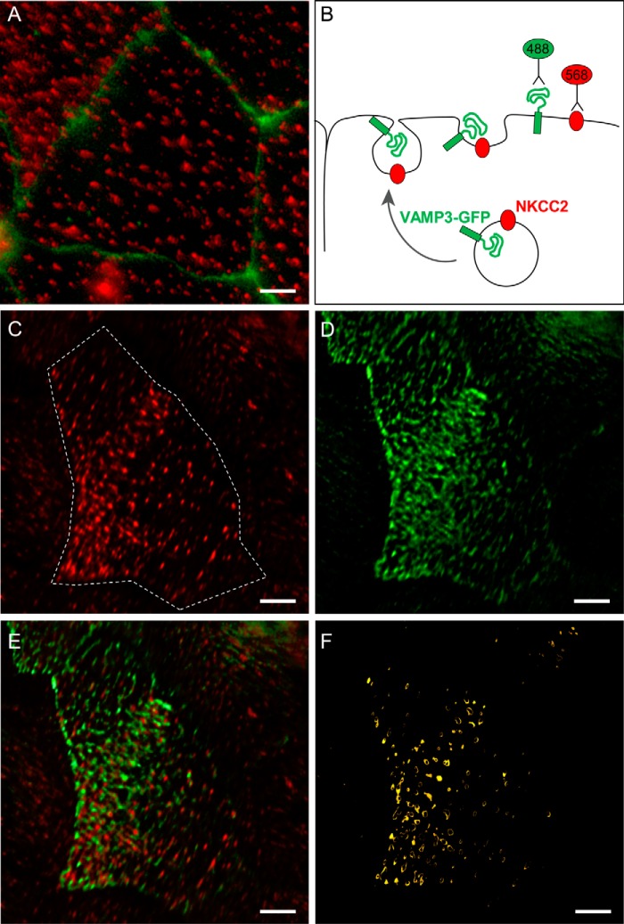
NKCC2 and VAMP3 co-localize at the apical surface of TALs. A, endogenous expression of NKCC2 in clusters at the apical surface of TAL primary cultures. Apical surface NKCC2 was detected with an antibody against an extracellular epitope applied to the apical side of non-permeabilized cultures (red). Individual cells are delimited by the tight junction marker ZO1 (green). B, schematic representation of the procedure followed to co-localize NKCC2 and VAMP3 at the apical surface of TAL primary cultures. The cells were transduced with VAMP3-GFP, which was detected at the apical surface with an anti-GFP antibody before permeabilizing. Endogenous NKCC2 was detected at the apical surface with the extracellular antibody. C, surface NKCC2 expression in TAL primary cultures (single cell indicated by the dashed line). D, surface VAMP3-GFP expression in apical clusters in TAL primary culture (same cell as in C). E, merged image showing co-localization of surface NKCC2 and surface VAMP3-GFP as indicated by the yellow color in some clusters. F, the apical clusters where surface NKCC2 and VAMP3 co-localize were identified by isolating the co-localizing pixels with a Mander's overlap coefficient ≥ 0.95 (n = 6). Scale bar, 1 μm.

