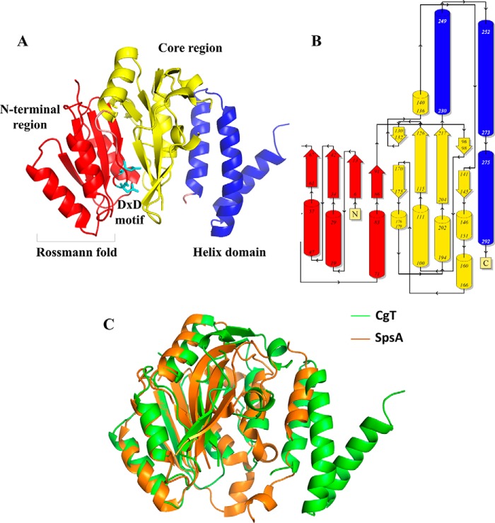
FIGURE 6.
Structure of the CgT monomer. The N-terminal region is colored red, core region is colored yellow, and the helix domain is colored blue. A, Rossmann-fold and DXD motif are labeled. Topology diagram of the CgT monomer was color coded as the structure. Arrows stand for strands, and columns represent helices (B). Superimposing of CgT with the best matched structure model of SpsA (C). CgT is shown in green, whereas SpsA is shown in orange.

