
FIGURE 3.
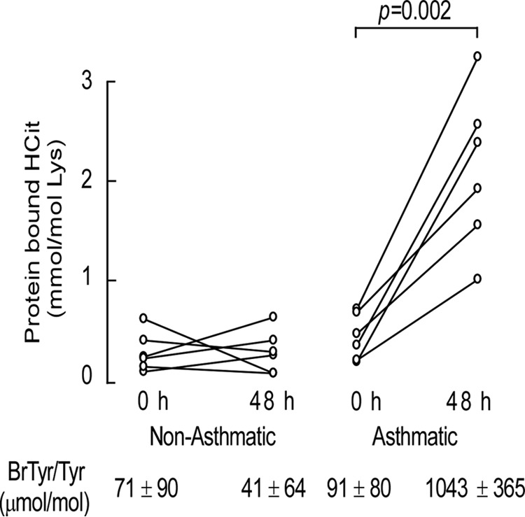
Protein-bound HCit is elevated in asthmatic subjects. Quantification of HCit in proteins recovered from non-asthmatic and asthmatic subjects at baseline and after segmental allergen challenge. Healthy control and allergic mild asthmatic subjects (n = 6 distinct subjects) underwent fiber optic bronchoscopy, and a specific segment of one lung was lavaged with normal saline to obtain a baseline sample (t = 0 h). The same specific segment in the contralateral lung was then exposed to allergen. Forty-eight hours later, fiber optic bronchoscopy was repeated on the allergen-exposed segment and lavaged with normal saline. Cells in the BAL were removed by centrifugation, and the levels of protein-bound HCit recovered in the supernatant at baseline and after segmental allergen challenge were then determined by LC/MS/MS. Abundance of protein-bound bromotyrosine (BrTyr) as an indicator of EPO-specific activity for each group is listed, as previously reported (14).