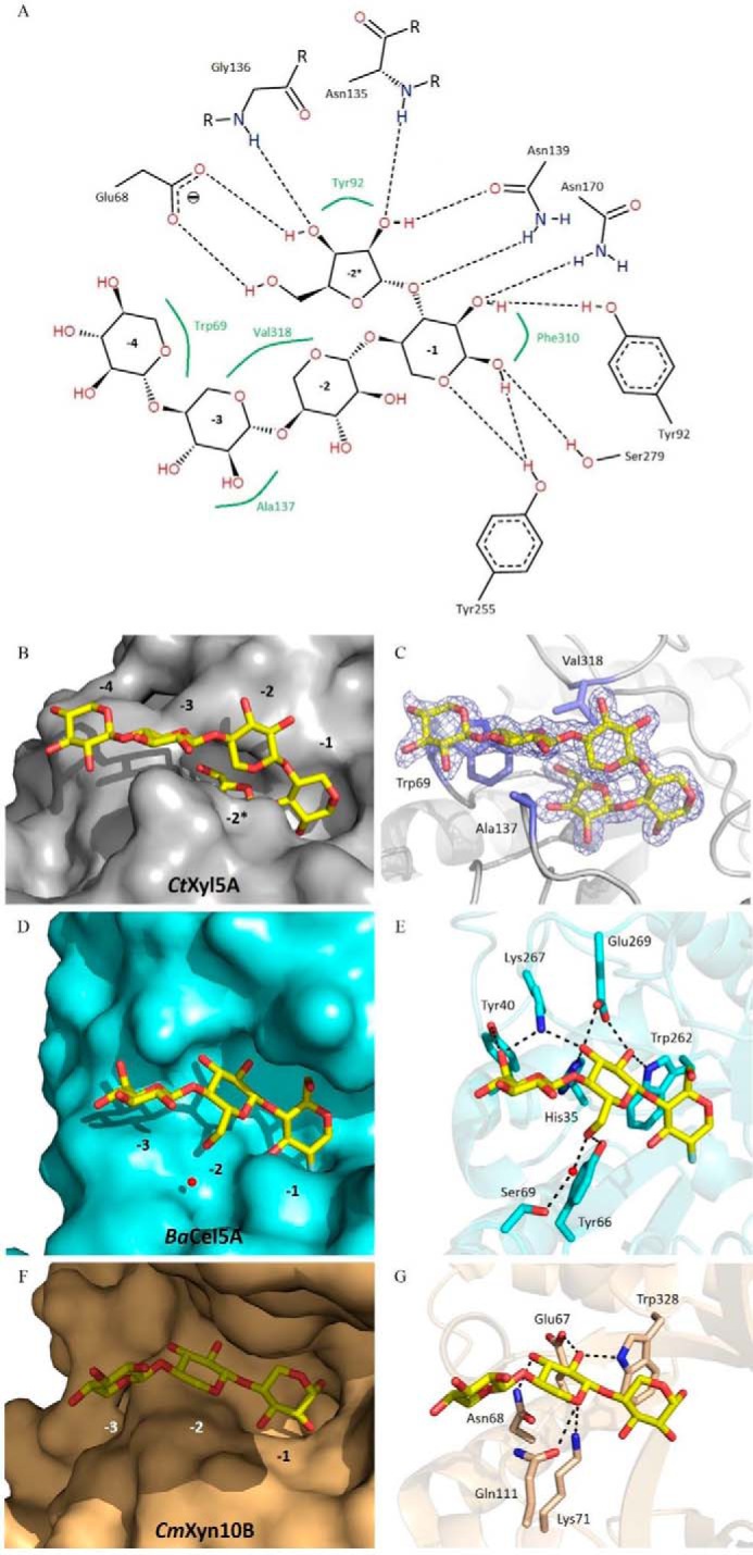
FIGURE 4.

Comparison of the ligand recognition at the distal negative subsites between CtGH5E279S-CBM6, the cellulase BaCel5A, and the xylanase GH10. A–C show CtGH5E279S-CBM6 is in complex with a pentasaccharide (β1,4-xylotetraose with an l-Araf linked α1,3 to the reducing end xylose). A, Poseview (40) representation highlighting the hydrogen bonding and the hydrophobic interactions that occur in the negative subsites. C, density of the ligand shown in blue is RefMac maximum-likelihood σA-weighted 2Fo − Fc at 1.5 σ. D and E display BaCel5A in complex with deoxy-2-fluoro-β-d-cellotrioside (PDB code 1qi2), and F and G show CmXyn10B in complex with a xylotriose (PDB code 1uqy). The ligand are represented as sticks. B, D, and F are surface representations (CtGH5E279S-CBM6 in gray, BaCel5A in cyan, and the xylanase GH10 in light brown). C, E, and G show the protein backbone as a cartoon representation with the interacting residues represented as sticks. The black dashes represent the hydrogen bonds.
