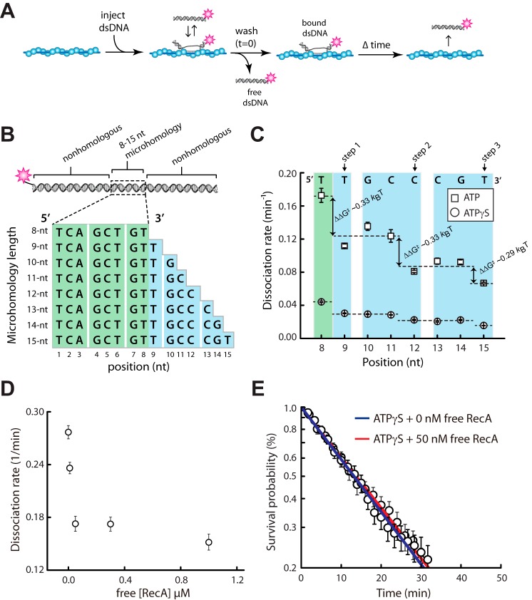
FIGURE 3.
Influence of ATP on base triplet recognition by RecA. A, experimental schematic for measuring dsDNA binding by the RecA presynaptic complex. B, schematic representation of the 70-bp dsDNA substrates used in the binding assays. The dsDNA is labeled at one end with a single Atto565 fluorophore and contains a single internal tract of microhomology ranging from 8 to 15 nt in length, as indicated, which are targeted to only one site on the M13 presynaptic ssDNA. C, dissociation rate data for oligonucleotides bearing different lengths of microhomology in reactions with either 1 mm ATP or 1 mm ATPγS; note that the data for experiments with ATPγS have been previously published (25) and are reproduced here for direct comparison with the ATP data. D, dissociation rate data for the dsDNA substrate bearing a single 8-bp tract of microhomology in the presence of varying concentrations of free RecA. E, dissociation rate data for reactions with the dsDNA substrate bearing a single 8-bp tract of microhomology in the presence of 1 mm ATPγS with and without 50 nm free RecA.

