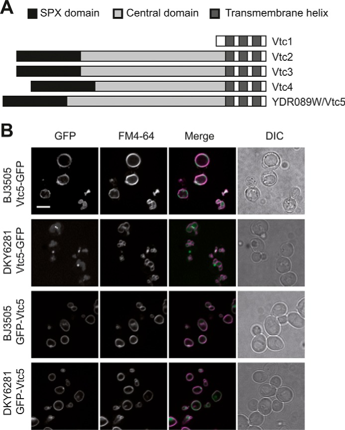
FIGURE 1.

Localization and topology of Vtc5. A, schematic representation of the predicted structural features of Vtc5 and comparison with previously known VTC subunits. Three transmembrane helices were predicted using the TMHMM server. B, Vtc5-GFP and GFP-Vtc5 localization was determined by confocal fluorescence microscopy in a pep4Δ strain (BJ3505) and in a strain expressing PEP4 (DKY6281). Cells were cultivated in YPD to logarithmic phase, and vacuoles were stained with the vital dye FM4-64. The N-terminally tagged version of Vtc5 (GFP-Vtc5) was expressed from the genomically integrated construct under control of the ADH1 promoter. The ADH1 promoter results in moderate overexpression. This explains the fusion of the vacuoles into a single larger organelle, which occurs as a consequence of enhanced polyP production (52). DIC, differential interference contrast. Scale bar, 5 μm.
