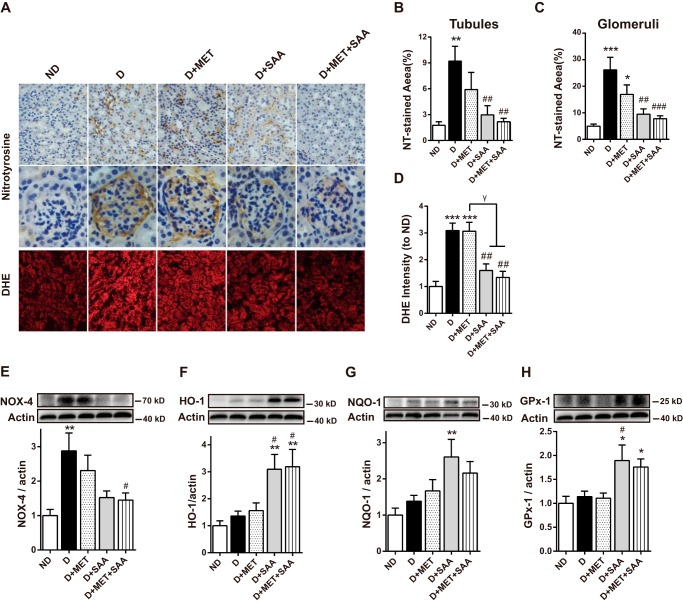
FIGURE 5.
Attenuation of oxidative stress by SAA or SAA in combination with MET was linked to the up-regulation of antioxidant enzymes. Oxidative stress in the kidney cortex was examined by NT immune staining in paraformaldehyde-fixed kidney and DHE fluorescent dye in kidney cryosections (A). The quantitative results in NT and DHE staining were assessed by Image-Pro Plus 6.0 (B–D). The protein level for NOX-4 was examined by Western blotting with its quantitation shown (E). Similarly, protein levels of several antioxidant enzymes, HO-1, NQO-1, and GPx-1 are shown (F–H). Data were mean ± S.E. n = 6–10 per group. *, p < 0.05; **, p < 0.01; ***, p < 0.001 versus ND; #, p < 0.05; ##, p < 0.01 versus D. γ, p < 0.05. ND, non-diabetic mice; D, diabetic mice; D+MET, diabetic mice treated with metformin; D+SAA, diabetic mice treated with SAA; D+MET+SAA, diabetic mice treated with SAA combined with metformin.

