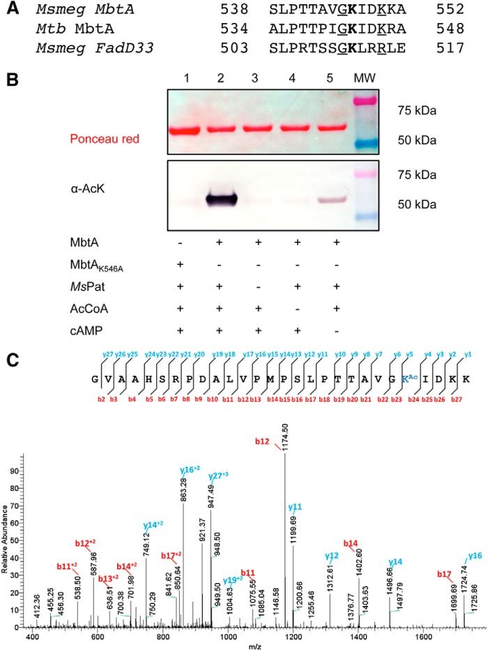
FIGURE 2.
Pat acetylates specifically MbtA on Lys546. A, partial alignments of MbtA proteins from M. smegmatis (MSMEG_4516) and M. tuberculosis (Rv2384) versus M. smegmatis FadD33 (MSMEG_2132). Acetylated lysines are indicated in bold type. Conserved residues and basic residues flanking acetylated sites are underlined. B, wild type MbtA or MbtA K546A mutant was incubated with multiple reaction components as indicated in the table. The samples were analyzed by Western blotting (bottom panel) with acetyl-lysine antibody (α-AcK), and total protein content was determined by Ponceau red (top panel). C, MbtA unique acetylation site was identified by MS/MS as Lys546. Shown in an MS/MS spectrum charged tryptic peptide from Ms MbtA (TTAVGKAcIDKK) bearing an acetylated lysine. The acetylated lysine is indicated as KAc.

