Figure 2.

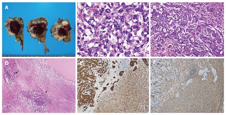
Pathology of the pancreatic tumor. A: Macroscopic appearance of the resected specimen. The tumor forms a poorly defined, whitish mass lesion measuring 60 mm in diameter. A large cyst filled with hemorrhagic material is present in the central portion of the tumor; B-C: Histology of the tumor. The major part of the tumor is composed of non-cohesive, pleomorphic tumor cells showing a sarcomatous appearance (B), whereas the minor part of the tumor is composed of irregularly shaped fused glandular structures, exhibiting the histology of moderately differentiated adenocarcinoma (C) [hematoxylin and eosin (HE) staining; original magnification, B: × 40, C: × 10]; D: Central part of the tumor. A cystic lesion filled with fluidic material (upper right) is surrounded by necrotic tumor tissue focally containing viable anaplastic carcinoma cells around the blood vessels (lower left/arrow) (HE staining; original magnification, × 4); E, F: Immunohistochemistry. Positive staining for pancytokeratin (AE1/AE3) is observed in both the adenocarcinoma component (E; left) and the sarcomatoid component (E; right), whereas positive vimentin staining is observed in the sarcomatoid component (F; right) and normal stroma but not in the adenocarcinoma component (F; left) (original magnification × 4).
