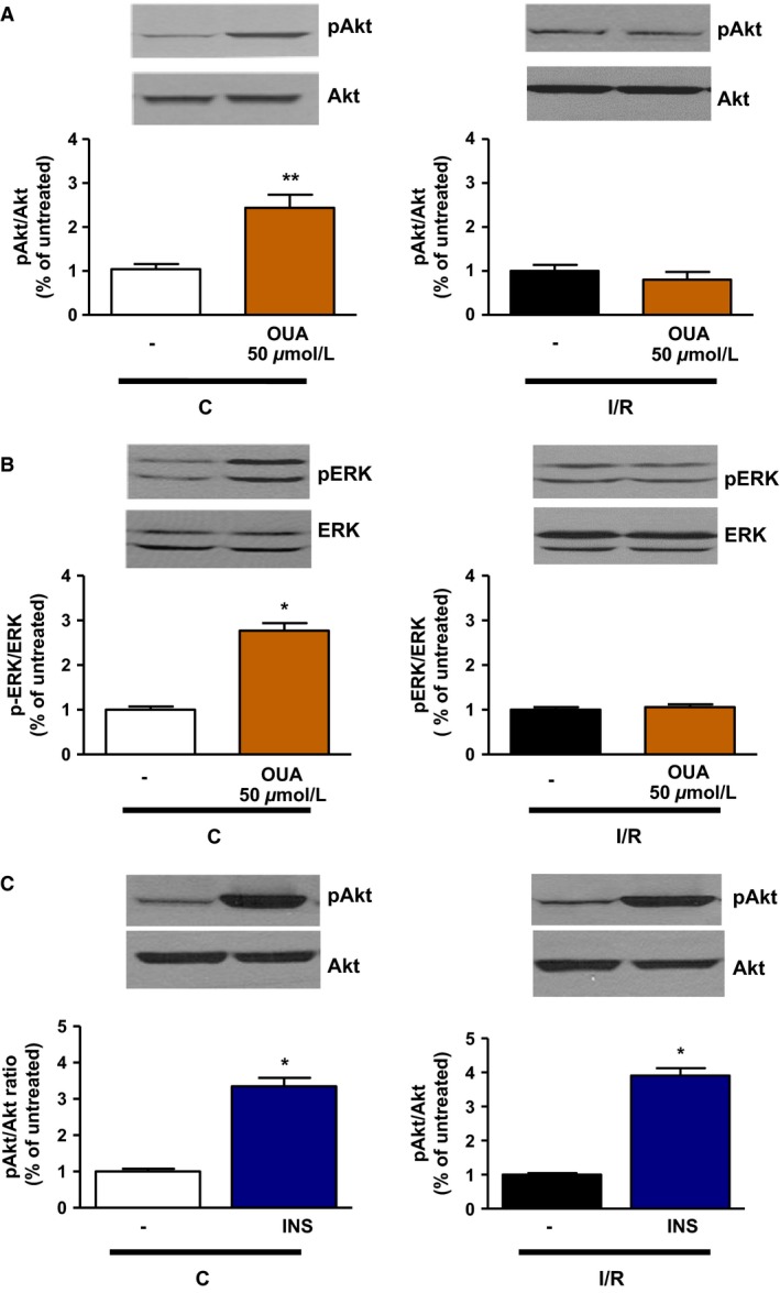
Figure 6.

Effect of ouabain 50 μmol/L on Akt and ERK phosphorylation in control and I/R hearts. Ouabain 50 μmol/L (A, B) or insulin 0.3 mU/mL (C) was added to the perfusate for 15 min after aerobic perfusion with Krebs–Henseleit buffer or following 30 min of ischemia and 30 min reperfusion, according to protocol B (Fig. 1). Crude homogenates were assayed for total and phosphorylated forms of Akt (A) and ERK (B). Upper panels: representative immunoblots. Lower panels: activation quantified as ratio of phosphorylated to total form of the indicated protein, normalized to C (left panels) or I/R (right panels). These ratios are normalized to one C (left panels) or one I/R (left panel) sample/gel, which was assigned the value of 1. Shown are means ± SEM from three to six independent experiments. *P < 0.05 and **P < 0.01 versus corresponding untreated condition.
