. 2016 Sep 30;28(5):624–628. doi: 10.5021/ad.2016.28.5.624

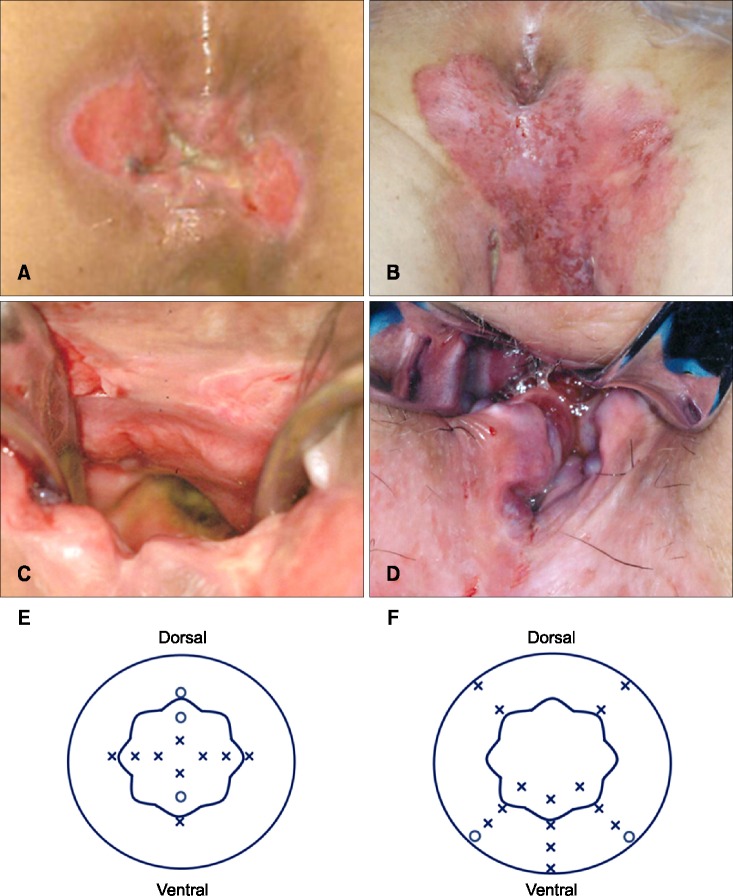
Fig. 1. Mapping biopsy of the anal canal. (A) A 69-year-old female with perianal extramammary Paget disease (EMPD) (the second patient). (B) The anal canal was sufficiently enlarged using a retractor in the jack-knife position under spinal anesthesia, and mapping biopsy of the anal canal was performed. (C) A scheme of the mapping biopsy of the anal canal. A total of 12 biopsies were performed (indicated by crosses and small circles): four on the dentate line, four 0.5 cm above the dentate line, and four 1.0 cm above the dentate line. The small circles demonstrate the sites that were positive for Paget cells. The large round circle indicates the anal verge, and the wavelike circle indicates the dentate line. (D) A 75-year-old female with vulvar EMPD (the sixth patient). The lesion involved the anal verge, and spread of Paget cells into the anal canal was highly suspected. (E) The anal canal was sufficiently enlarged using a retractor in the jack-knife position under spinal anesthesia. The spread of Paget cells into the anal canal were particularly suspected in the vertebral side. (F) A scheme of the mapping biopsy of the anal canal. A total of 16 biopsies were performed: five on the anal verge, three 10 mm above the anal verge, five on the dentate line, and three 10 mm above the dentate line. The small circles demonstrate the sites that were positive for Paget cells.

Fig. 1