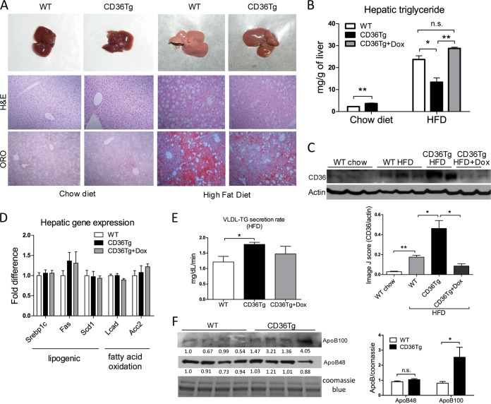
FIG 3.
CD36Tg mice were protected from HFD-induced hepatic steatosis. (A) Gross appearance (top) and histology (bottom) (H&E and Oil Red O [ORO] staining) of livers of mice maintained on a chow diet (left) and mice that had been fed an HFD for 19 weeks (right). (B) Hepatic triglyceride levels in mice maintained on a chow diet and mice that had been fed an HFD for 19 weeks in the absence or presence of Dox. n = 5. (C) Expression of liver CD36 as measured by Western blotting. Densitometric quantifications of the blots are shown below. (D) The hepatic expression of genes involved in lipogenesis and fatty acid oxidation in HFD-fed mice was measured by real-time PCR. n = 5. (E) VLDL-triglyceride (TG) secretion rate in HFD-fed WT, CD36Tg, and CD36Tg-plus-Dox mice. n = 4. (F) The serum levels of ApoB100 and ApoB48 were measured by Western blotting. The serum samples were run on two parallel gels; one was used for Coomassie blue staining as a loading control, and the second gel was used for ApoB100/48 Western blotting. The densitometric intensity was normalized to the Coomassie blue staining. The densitometric scores for individual lanes are shown. On the right is shown statistical analysis of the densitometric quantification. *, P < 0.05; **, P < 0.01; n.s., not statistically significant. The data are presented as means and SEM.

