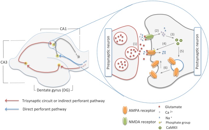
Figure 1.
The two main pathways to the CA1 area of the hippocampus on the left, and early-phase NMDA dependent-LTP on the right. The red arrows in the picture on the left show the trisynaptic circuit of the hippocampus, where multimodal sensory and spatial information coming from the entorhinal cortex (EC) is relayed to the CA1 area following this route: EC–DG–CA3–CA1. In blue, we illustrated the direct perforant pathway, which directly connects the EC to the CA1 region. On the picture in right, we show an illustration of the early-phase LTP. Here, (1) glutamate from the presynaptic neuron is released into the synaptic cleft. (2) This neurotransmitter reaches ionic channels of the postsynaptic cell causing depolarization of this neuron by the influx on sodium and calcium cations. (3) Calcium, in its turn, activates CaMKII that (4) phosphorylates ionic channels in the PSDs and (5, 6) induces the addition of AMPA receptors to the postsynaptic membrane, increasing synaptic efficiency.

