Figure 5.
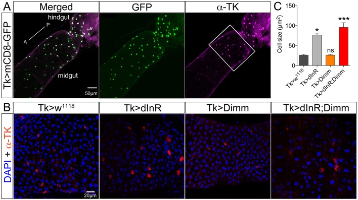
Tachykinin (TK) expressing enteroendocrine (EE) cells in adult midgut are Dimm negative and grow after ectopic expression of dInR and Dimm/dInR. (A) TK immunolabeling (magenta) in posterior midgut and anterior hindgut is colocalized in EEs with GFP expression driven by Tk-Gal4. (B) Posterior region of midgut (white bracket in A) was monitored after manipulations. Nuclei were marked with DAPI (blue) and EE cells were stained with anti-TK antibody (red). Cell sizes increased significantly after dInR expression and Dimm/dInR expression, compared to control, and Dimm alone. (C) Cell body sizes after dInR, Dimm, and Dimm/dInR expression compared to controls. Data are presented as means ± S.E.M, n = 6–9 flies for each genotype from three independent crosses (*p < 0.05, ***p < 0.001 as assessed by unpaired Students' t-test or non-parametric Mann Whitney test when data was not normally distributed). Scale bar = 50 μm in (A), 20 μm in (B).

